















































Quino
Liniers
Tute
Album
Akg
Magnum Photos
THE NEW YORK TIMES
Fotoarena
Researchers
Bridgeman
Granger
Verger
Minden
Interfoto
Science Source
Zuma
Universal
Everett
Nature
Newscom
DPA
Sipa
Glasshouse Images
Deposit
Quadrinhos
Saulo
Rita
Reuters Pictures
Science Photo Library
ImageBROKER
Angeli
Cagle Cartoons
Fabiane Langona
Caia Image
Almir Almeida
Márcio Pannunzio
María Verónica
André Barroso
PA Images
Ani Ganzala
Caloi
Ingran
British Museum
Allan Sieber
Angel Boligán
Adão Iturrusgarai
Claudia Liz
Cassio Vasconcellos
Flávio Cannalonga
Alamy
SPP
Robert Harding
TopFoto
Nando Motta
Ciro Fernandes
African Pictures

Busque também em nossas outras coleções:

Tipo de arquivo:

Tipo do arquivo:

Tipo da licença:

Orientação:

Total de Resultados: 62.869

Página 1 de 629

UDK844446
DC
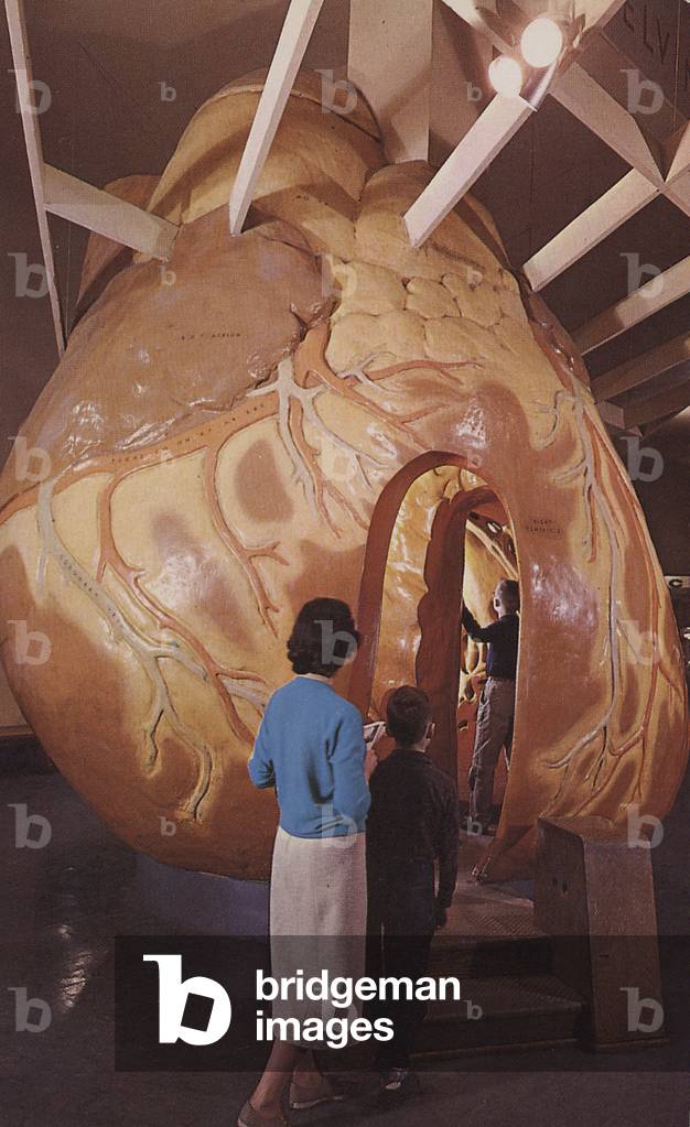
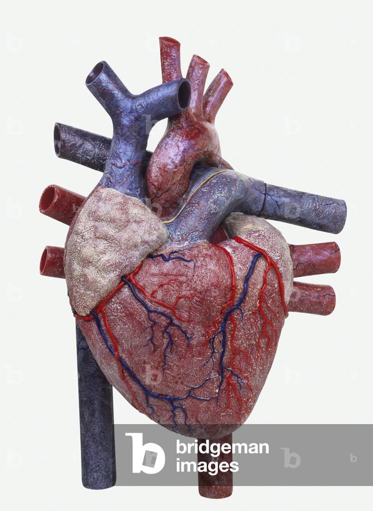
SIC743619 Museum visitors walk through a large model of the human heart and its arteries at the Museum of Science and Industry in Chicago, Illinois.
DC
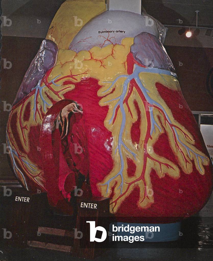
SIC743605 A cardiovascular exhibit housed at the Museum of Science and Industry in Chicago, Illinois, allows museum visitors to enter into a large model of a human heart and its arteries.
DC
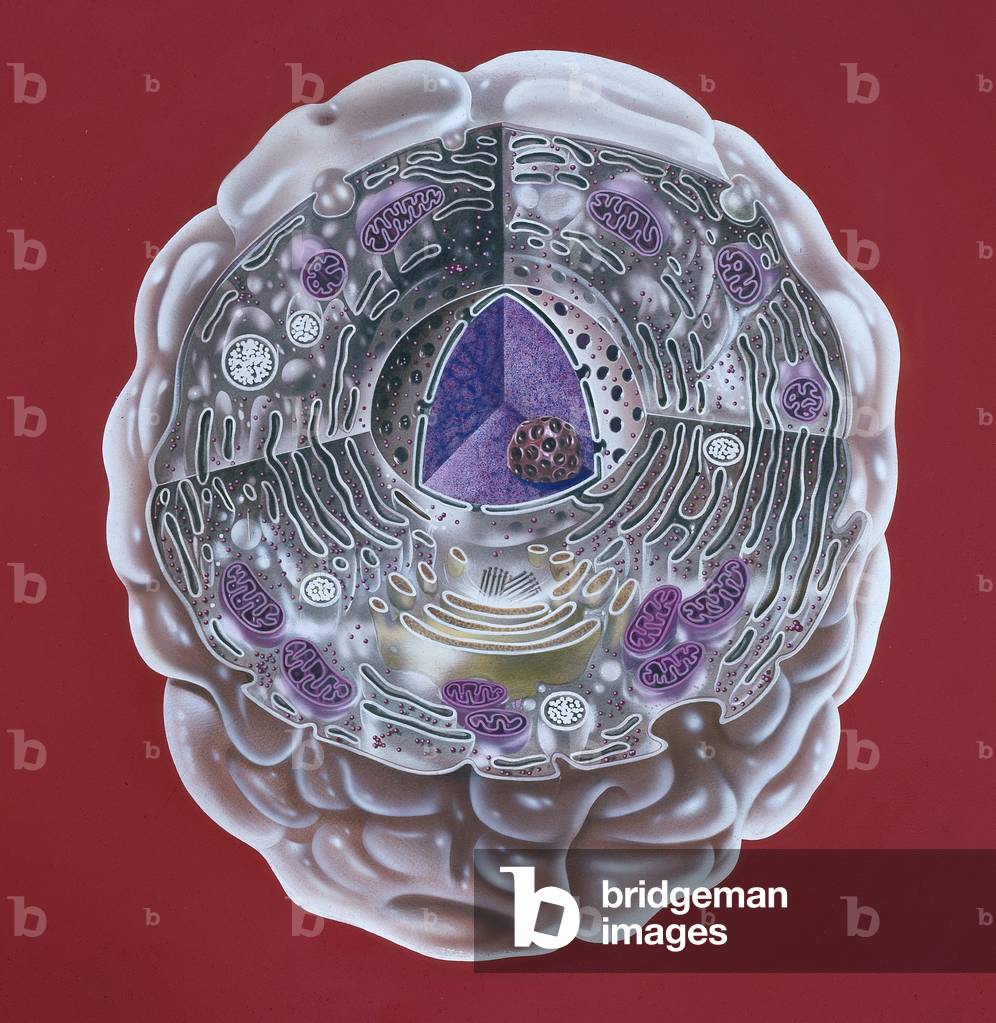
NHM1452569 Human cell, Illustration of a highly magnified section through a human cell. Page 8 from Human Biology, 1977. - Biology
DC
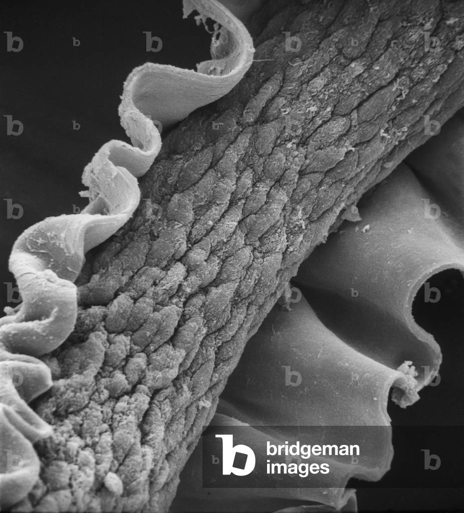
NHM1456175 Human hair, Scanning electron microscope (SEM) image showing a human hair with the cuticle reflexed. - Animal
DC
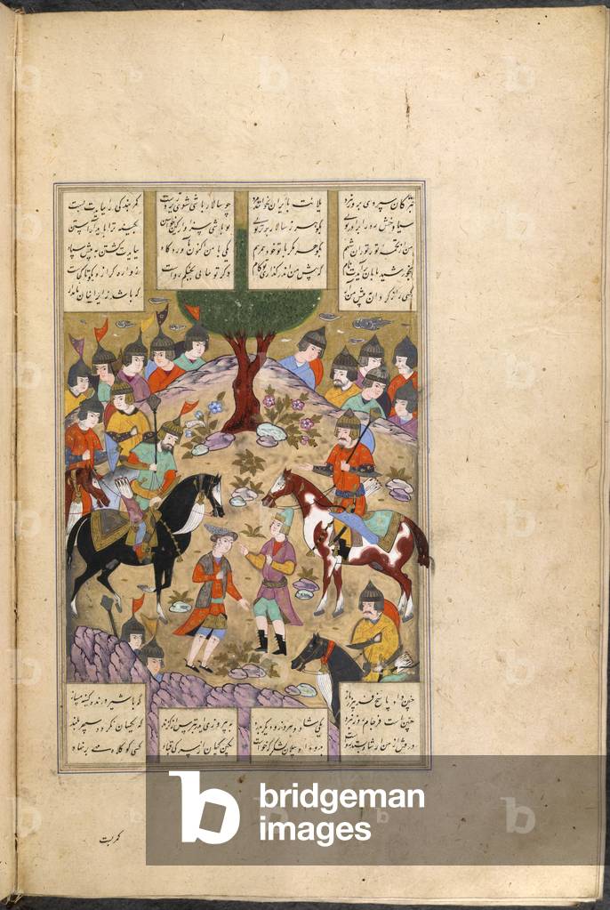
BL3276444 Shahnama of Firdawsi, with 63 miniatures.Illustrator: (Anon)1604Language: PersianSource/Shelfmark: I.O. ISLAMIC 966, f.212vParley between Gudarz and Human. 24.5 by 15.5 cm.Image taken from Shahnama of Firdawsi, with 63 miniatures.Originally published/produced in 1604.
DC
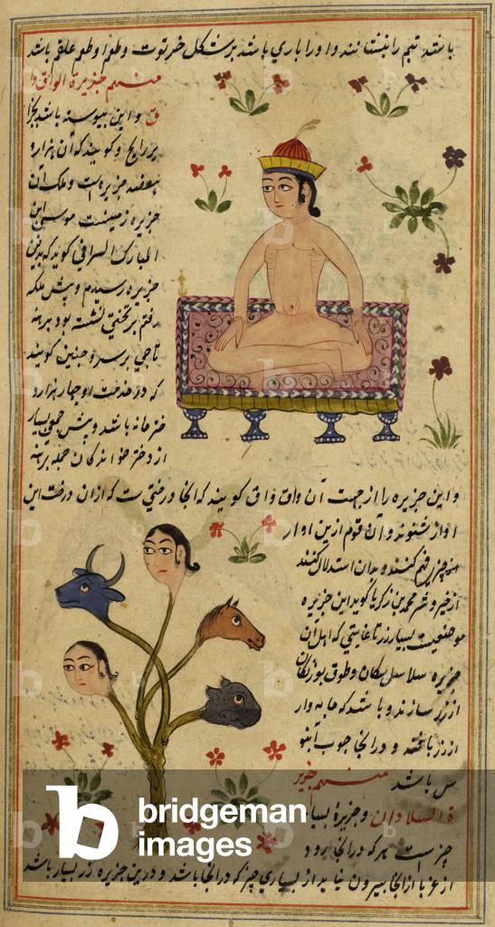
BL3306659 Aja'ib-almakhlukat wa ghara'ib almaujudatLanguage: Persian Source/Shelfmark: I.O. ISLAMIC 1919, f.50vThis is an undated translation from Arabic into Persian of the first part of 'Aja'ib-almakhlukat wa ghara'ib almaujudat' by the thirteenth century sage al-Kazwini (sometimes rendered as al-Qazwini).
DC
UIG531220
DC
UIG534713
DC
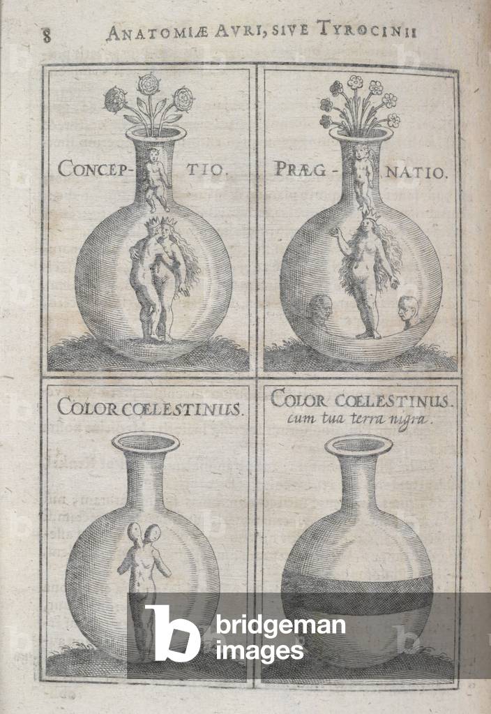
BL3276847 J. D. Mylii ... Anatomia Auri, sive Tyrocinium medFrancofurti, 1628Source/Shelfmark: 1033.l.6.(1), 8Various stages of human development showing conception and pregnancy.Image taken from J. D. Mylii Anatomia Auri, sive Tyrocinium medicochymicum, continens in se partes quinque: Quarum I. tradit concordantiam et harmoniam solis coelestis cum auro terrestri, item auri definitionem et confusam multorum physicorum de auro opinionem. II. agit de medicinis aureis et receptis antiquorum ac recentium Medicorum III. tractat de auri potabilis praeparatione IV. exhibet usum medicinalem auri potabilis V. demonstrat ideam Lapidis Philosophici in duodecim figuris.Originally published/produced in Francofurti, 1628.
DC
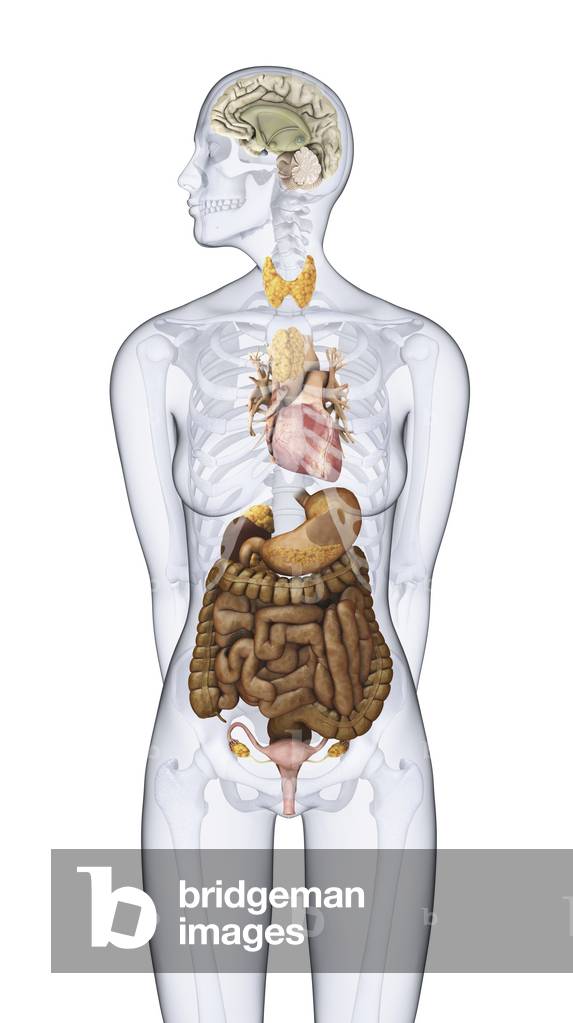
PRX997669 From ‘Bilderbuch für Kinder’ (Picture Book for Children) 1790-1830, published by Friedrich Justin Bertuch (1747-1822) Lage der Eingeweide im menschlichen KörperDie geöffnete Brust- und Bauchhöhle nach Wegnahme mehrerer Eingeweide, die geöffnete Brust- und Bauchhöhle in einer anderen Ansicht, Anatomie
DC
UIG534705
DC
UIG534708
DC
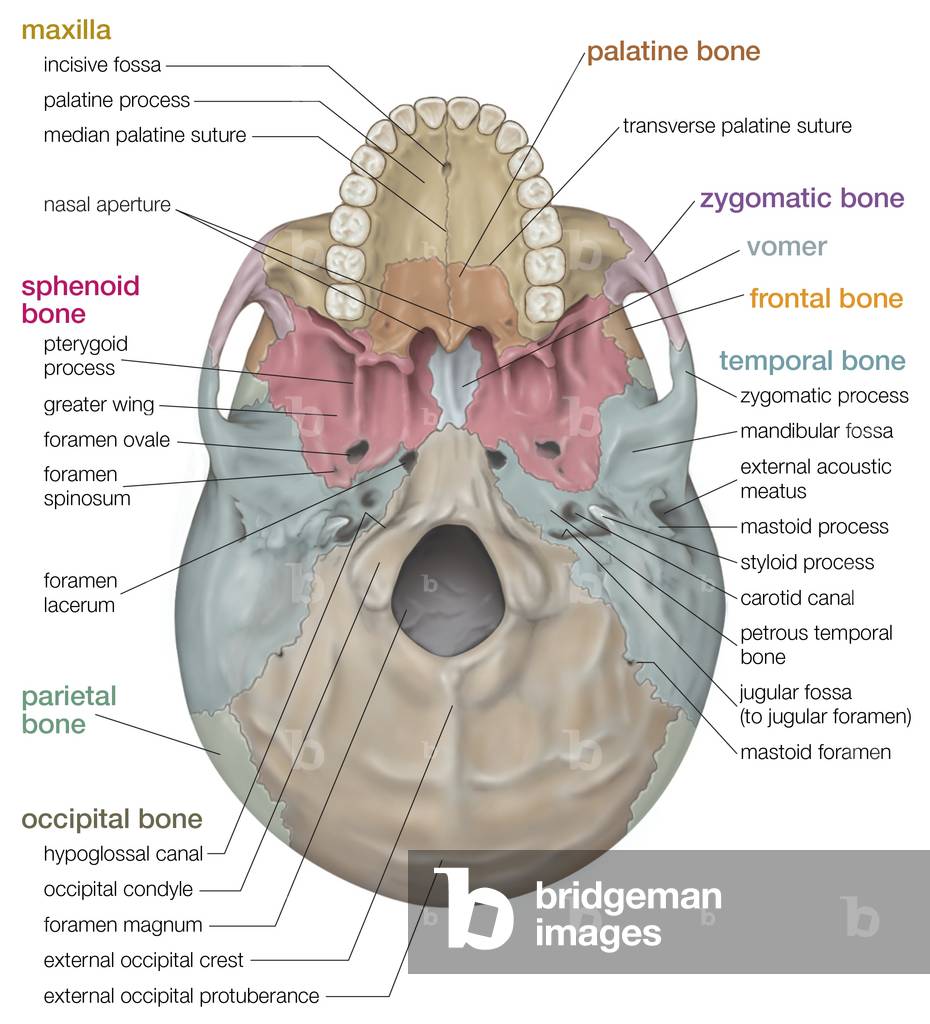
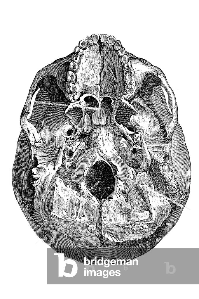
UIG535189 Inferior view of the human skull.
DC
UDK844263
DC
UIG535373
DC
UIG535218 The bony framework of the human head and neck.
DC
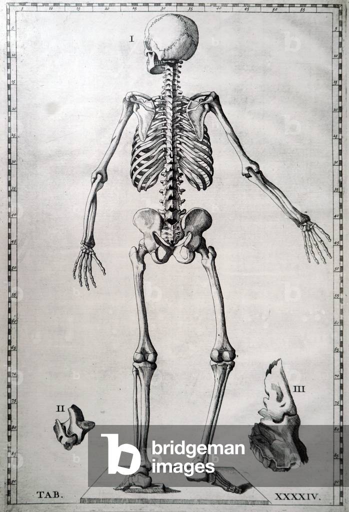
UIG5308004 Print of a human skeleton by Bartolomeo Eustachi(d.1574) one of the founders of the science of human anatomy. Dated 18th century.
DC
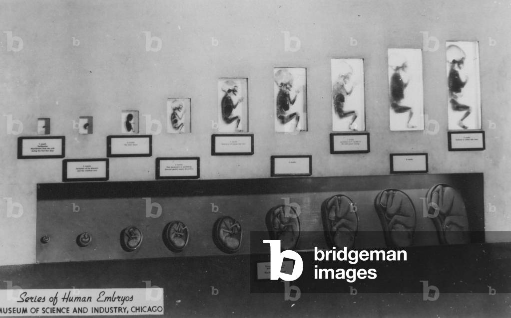
SIC743590 An exhibit housed at the Museum of Science and Industry in Chicago, Illinois shows a series of human embryos in various stages of development.
DC
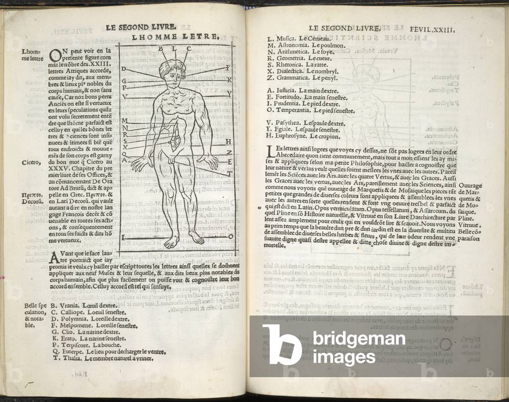
BL3274615 Champ Fleury. Auquel est contenu Lart & Science deAuthor: Tory, Geofroy / Geofroy Tory: Paris, 1529Language: FrenchSource/Shelfmark: 60.e.14, e4v-e5Diagram of a human figure.Image taken from Champ Fleury. Auquel est contenu Lart & Science de la deue & vraye Proportiõ des Lettres Attiques, quõ dit autremet Lettres Antiques, & vulgairement Lettres Romaines proportionees selon le Corps & Visage humain. Few MS. notes.Originally published/produced in Geofroy Tory: Paris, 1529.
DC
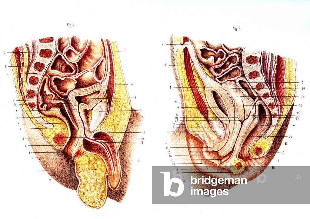
JAB4119239 Newborn male and female reproductive organs 1866 cutaway computer artwork comparing the anatomy of the reproductive system of a newborn boy (left) and girl (right) In a newborn boy the testes are held within the body and descend at puberty Anatomy of the pelvic organs - Fig 1: boys' organs new ne, Fig 2: girls' organs new nee, 1866
DC
UIG5309305 Engraving depicting a human heart. Dated 18th century01/02/1764
DC
UIG534701
DC
UIG534533
DC
XEE4156429 Human Zoo - Sudan in Paris (ethnographic exhibition of West Africa at Champ de mars (Paris), 1895. drawing by Pierre Georges Jeanniot (1848-1934) for “” Le Rire”” of 28 September 1895
RF
UIG805920
DC
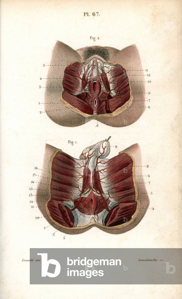
FLO4672956 Circulatory system of the genitals. Lithograph of Anneshower, based on a drawing by Leveille, in Petity Atlas complet d'Anatomie descriptive du Corps Humain, by Dr. Joseph Nicolas Masse, published by Mequignon Marvis, Paris (France), 1864.
DC
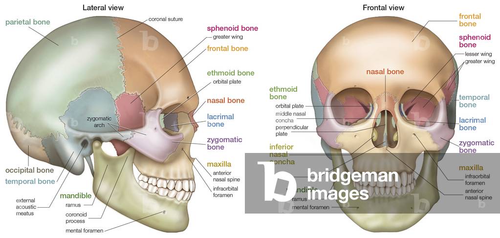
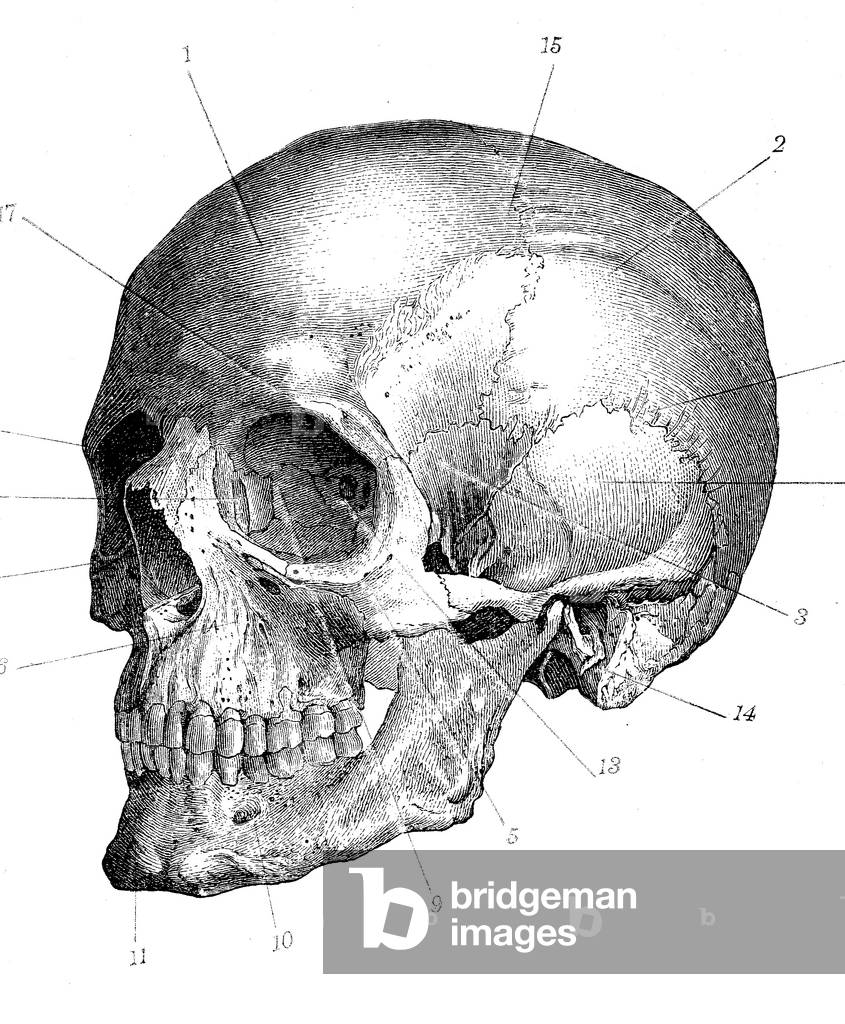
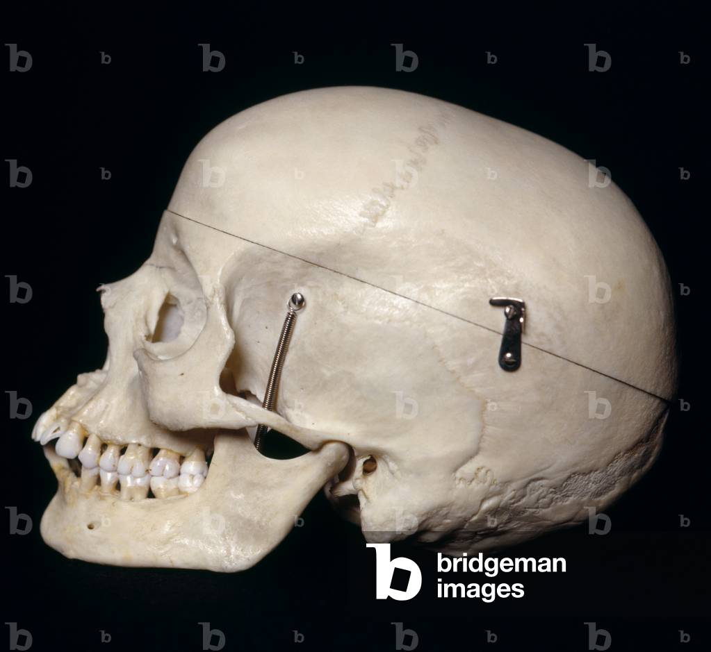
UIG535186 (Left) Lateral and (right) frontal views of the human skull.
DC
UDK5430048
DC
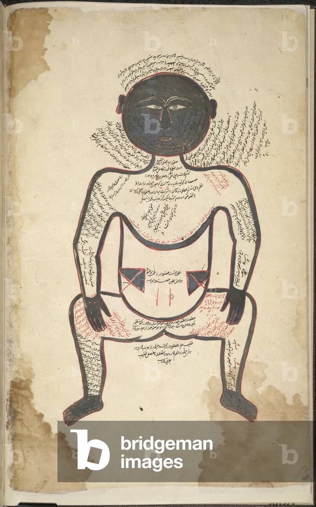
BL3261972 Tashrih bi al-Taswir.Author: Mansur bin Muhammad / Iran, 1672Language: PersianSource/Shelfmark: I.O. ISLAMIC 1379, f.11vThe human body illustrating the muscular system. The face and outlines are blackened silver. A miniature painting from a seventeenth century manuscript of Tashrih bi al-Taswir, a treatise on human anatomy by Mansur bin Muhammad.Image taken from Tashrih bi al-Taswir.Originally published/produced in Iran, 1672.
DC
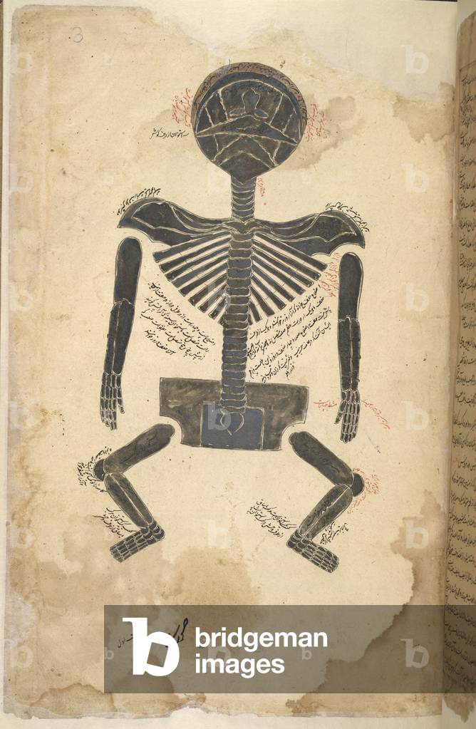
BL3261975 Tashrih bi al-Taswir.Author: Mansur bin Muhammmad / Iran, 1672Language: PersianSource/Shelfmark: I.O. ISLAMIC 1379, f.3The back view of a human skeleton. The tinting is in silver, which has blackened. A miniature painting from a seventeenth century manuscript of Tashrih bi al-Taswir, a treatise on human anatomy by Mansur bin Muhammad.Image taken from Tashrih bi al-Taswir.Originally published/produced in Iran, 1672.
DC
PRX997587 From ‘Bilderbuch für Kinder’ (Picture Book for Children) 1790-1830, published by Friedrich Justin Bertuch (1747-1822) Menschenhaut und BlutMenschenhaut, Menschenblut, Humanmedizin, Mensch
DC
HLR5891691
DC
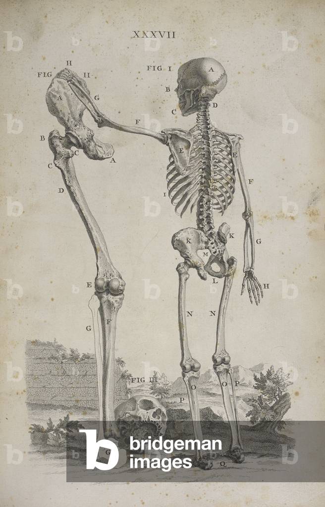
BL3314070 Osteographia, or the Anatomy of the bones.Author: Cheselden, William / London, 1733Source/Shelfmark: 458.g.1 plate XXXVII
DC
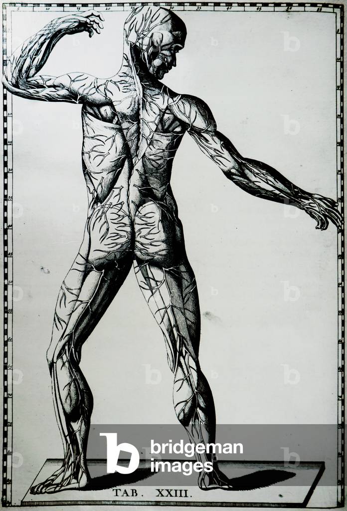
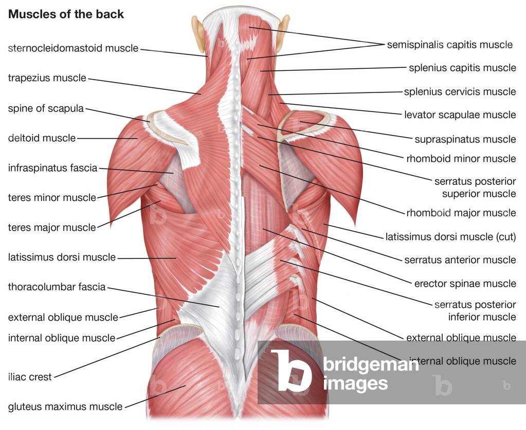
FLO4591171 Anatomy of human musculature from the back showing outer muscles. Copperplate engraving by Milton from Abraham Rees' Cyclopedia or Universal Dictionary of Arts, Sciences and Literature, Longman, Hurst, Rees, Orme and Brown, London, 1820.
DC
UIG531473
DC
BAL67863
DC
HSC257649
DC
BAL33592 by Jose Clemente Orozco
DC
UIG5321771 human skull bones, digital improved reproduction from an original print from the year 1881
DC
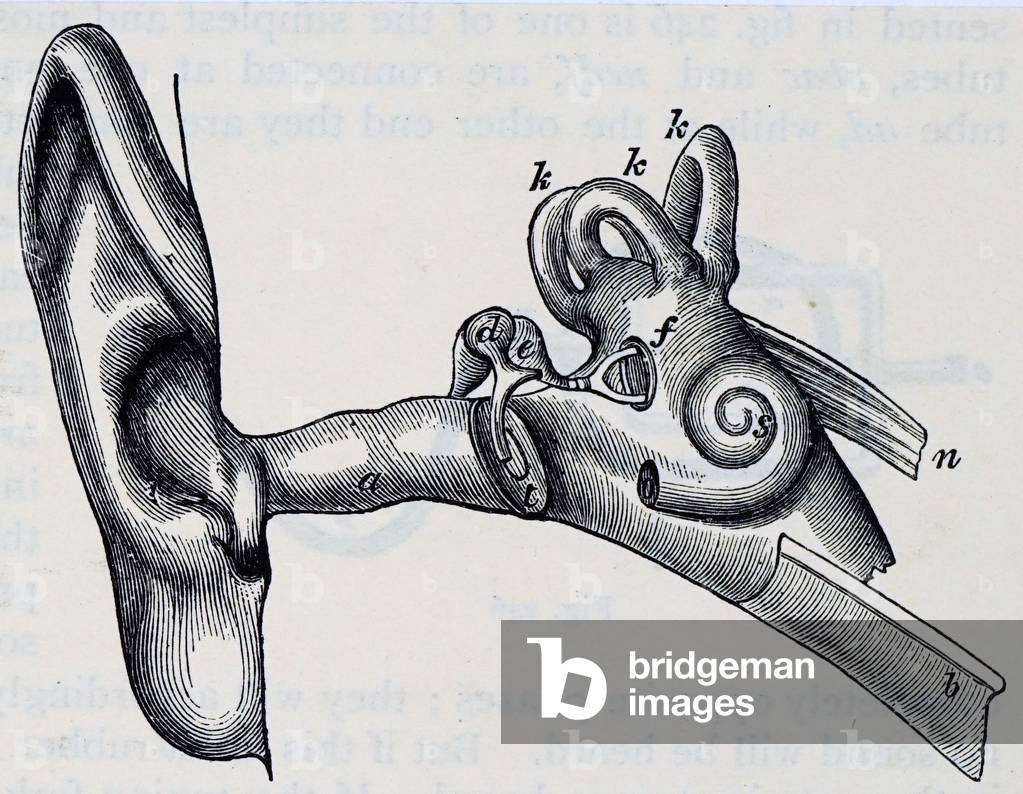
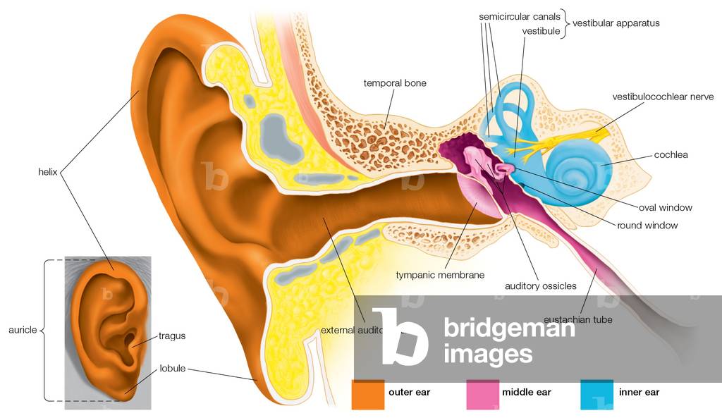
UIG5309259 Diagram of the human ear. A: auditory passage. T: Tympanum. D: hammer. E: anvil. F: stirrup and oral window. B: eustachian tube. K: semi-circular canals. S: cochlea. Dated 20th century01/02/1906
DC
UDK844254
DC
UDK844231
DC
UDK844258
DC
BL3283502 DRAWINGS, in Indian ink, illustrative of Capt. Cook's first voyage, 1768 -1770, chiefly relating to Otaheite and New Zealand, by A. Buchan, John F. Miller, and others.Illustrator: Buchan, A; Parkinson, S; Miller J F1768-1770Source/Shelfmark: Add. 15508, no.48
DC
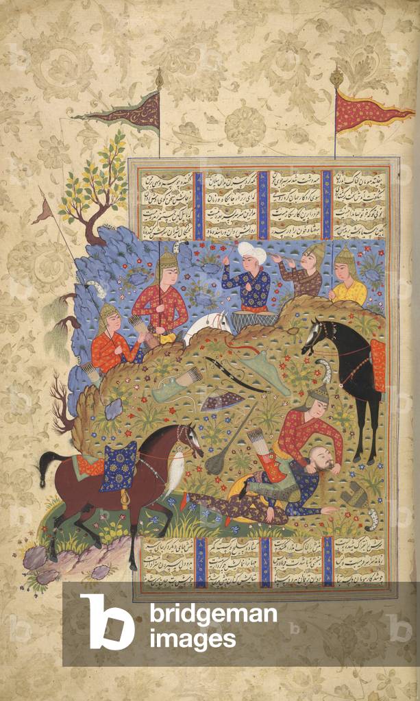
BL3275847 Shahnama.Author: Firdawsi / Kazvin, Iran, c.1590-1595Language: PersianSource/Shelfmark: Add. 27257, f.206Bizhan killing Human. Opaque watercolour. Safavid/Qazwin style.Image taken from Shahnama.Originally published/produced in Kazvin, Iran, c.1590-1595.
DC
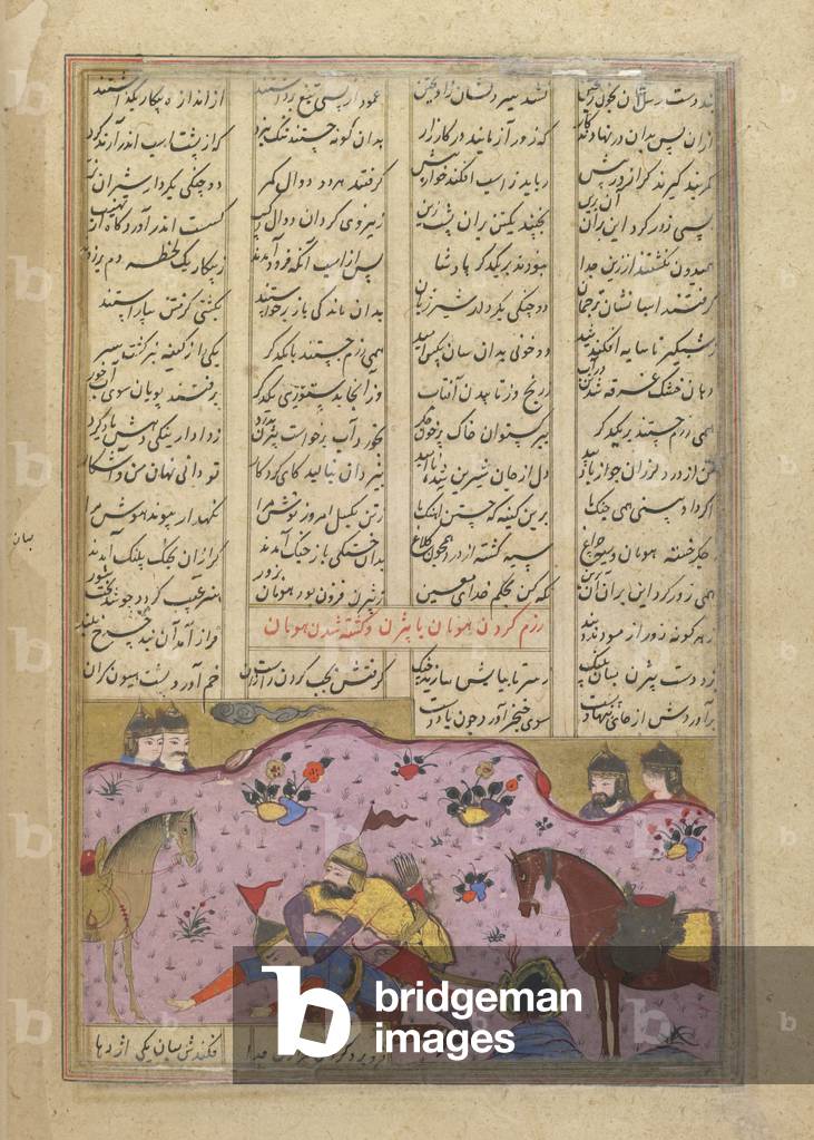
BL3275968 Shahnama of Firdawsi, with 49 miniatures. Opaque wIllustrator: [Anon]1590-1600Language: PersianSource/Shelfmark: I.O. ISLAMIC 3254, f.216vHuman slain by Bizhan. 8.5 by 14.5 cm.Image taken from Shahnama of Firdawsi, with 49 miniatures. Opaque watercolour. Safavid/Isfahan style.Originally published/produced in 1590-1600.
DC
UIG534551
DC
PIX4675754 Neanderthal meets Homo Sapiens - Neanderthal meets Homo Sapiens - A Neanderthal woman (Homo neanderthalensis), on the left, meets a Homo Sapiens. The two species probably crossed when Homo Sapiens arrived in Europe 42,000 years ago. A Neanderthal woman (Homo neanderthalensis), to the left, meets an Homo Sapiens. Both have probably met when Homo Sapiens arrived in Europe 42,000 years ago
DC
UIG534858
DC
PIX4639956 Mars Exploration - Mars Exploration - Illustration: Astronauts drill the surface of Mars to find water. Astronauts drill for water on the Red Planet.
DC
PIX4650338 Traces of the genetic mutation- Artist's view - Tracking genetic mutations - Artist vie
DC
FLO4590679 Anatomy of human internal organs from the front showing heart, stomach, intestines, kidneys and bladder. Copperplate engraving by Milton from Abraham Rees' Cyclopedia or Universal Dictionary of Arts, Sciences and Literature, Longman, Hurst, Rees, Orme and Brown, London, 1820.
DC
FLO4591048 Anatomy of human musculature from the back showing inner muscles. Copperplate engraving by Milton from Abraham Rees' Cyclopedia or Universal Dictionary of Arts, Sciences and Literature, Longman, Hurst, Rees, Orme and Brown, London, 1820.
DC
PIX4650398 Teleportation - Artist's View
DC
UIG531218
DC
PIX4639670 Inhabited Mission on Mars -Illustration - Astronauts on Mars - Illustratio
DC
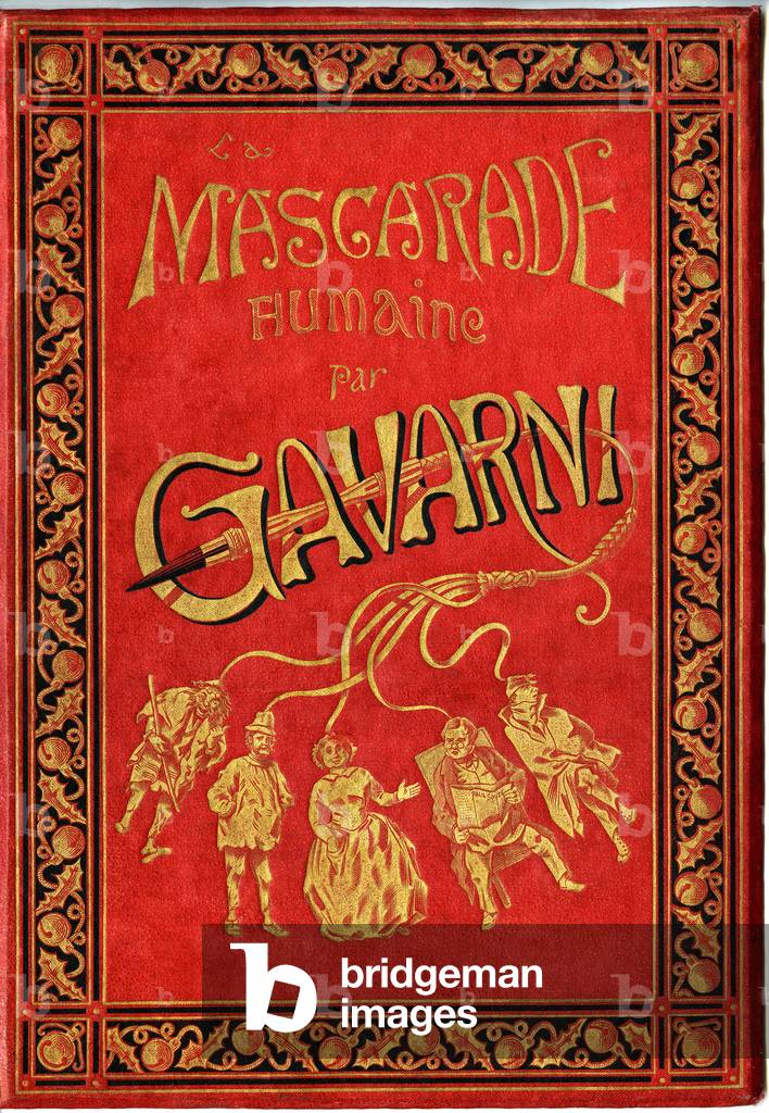
PCT4297613 Cover of the book “” The Human Masquerade””, caricatures by Gavarni (Sulpic-Guillaume Chevalier 1804-1866 dit Paul Gavarni). Editions Calmann Levy 1881, cardboard by Paul Souze, binding and gilding C. Magnier - characters at the end of a swift (whip): “” who likes chatie well””” -
DC
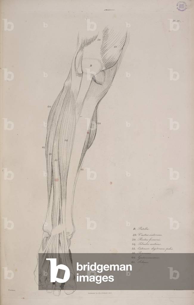
BL3298960 Anatomical studies of the bone and muscles for the use of artists. Author: Flaxman, John / London: M. A. Nattali, 1833. Source/Shelfmark: AX3/0118, plate 16
DC
UIG534866
DC
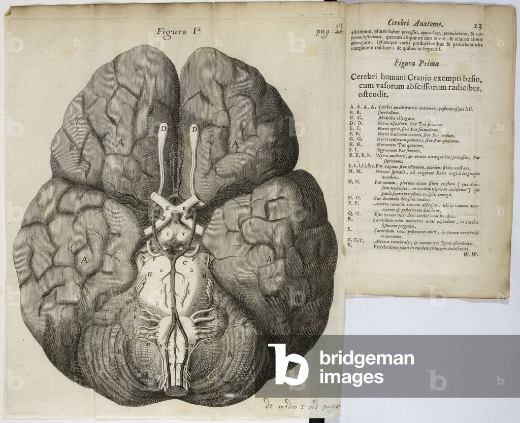
BL3295676 Cerebri anatome, cui accessit nervorum descriptio et usus.Author: Willis, Thomas / Londini, 1664Source/Shelfmark: 1607/109, page 13 and opposite
DC
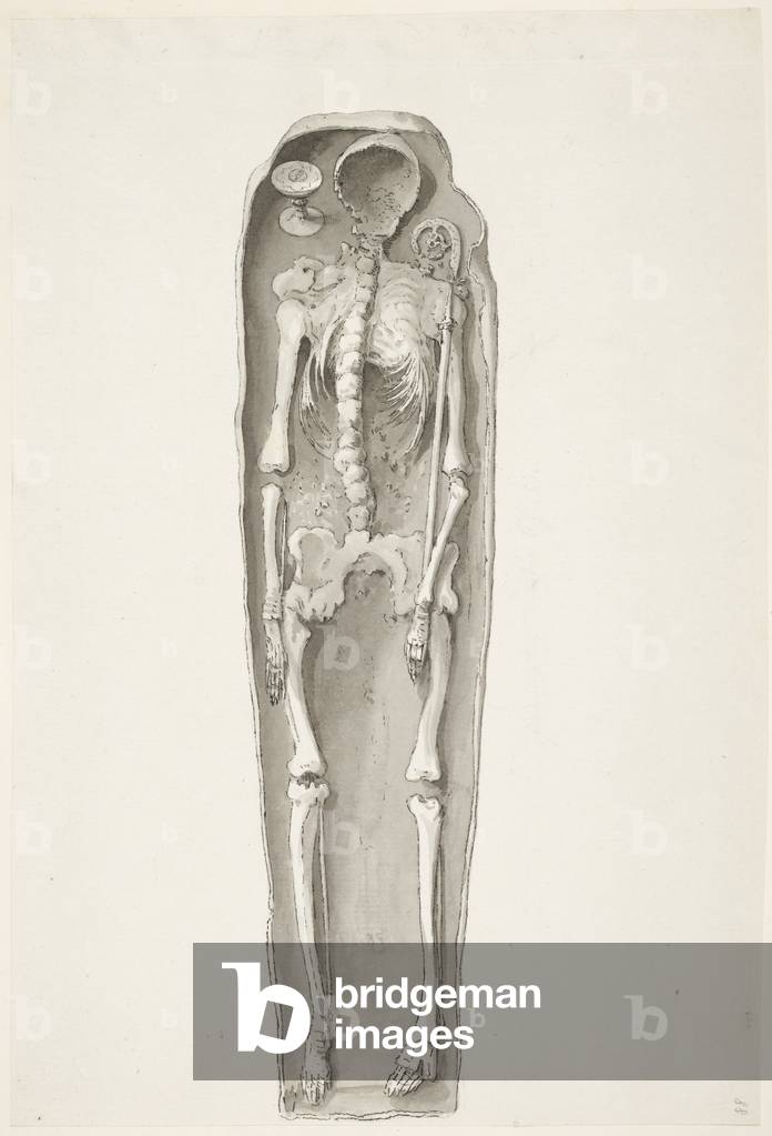
BL3285542 Source/Shelfmark: Add. 15541 f.88From a collection of drawings in pencil or Indian ink, representingviews, buildings, and objects of antiquity, or curiosity in variouscounties of' England; drawn in the latter part of the last century, bySamuel Henry Grimm, for the Very Rev. Sir Richard Kaye, Bart.,Dean of Lincoln.
DC
FLO4590759 Anatomy of human internal organs from the back showing stomach, liver, intestines, gallbladder, etc. Copperplate engraving by Milton from Abraham Rees' Cyclopedia or Universal Dictionary of Arts, Sciences and Literature, Longman, Hurst, Rees, Orme and Brown, London, 1820.
DC
FLO4590988 Anatomy of human musculature. Copperplate engraving by Milton from Abraham Rees' Cyclopedia or Universal Dictionary of Arts, Sciences and Literature, Longman, Hurst, Rees, Orme and Brown, London, 1820.
DC
FLO4591432 Anatomy of human musculature showing outer muscles from the front. Copperplate engraving by Milton from Abraham Rees' Cyclopedia or Universal Dictionary of Arts, Sciences and Literature, Longman, Hurst, Rees, Orme and Brown, London, 1820.
DC
FLO4594255 Anatomy of human internal organs from the back showing spine, lungs, liver, intestines, gallbladder, etc. Copperplate engraving by Milton from Abraham Rees' Cyclopedia or Universal Dictionary of Arts, Sciences and Literature, Longman, Hurst, Rees, Orme and Brown, London, 1820.
DC
FLO4591280 Anatomy of human musculature from the front showing inner muscles. Copperplate engraving by Milton from Abraham Rees' Cyclopedia or Universal Dictionary of Arts, Sciences and Literature, Longman, Hurst, Rees, Orme and Brown, London, 1820.
DC
UIG534838
DC
PRX1000362 Engraver:Johann Carl BockFrom ‘Die Säugthiere in Abbildungen nach der Natur mit Beschreibungen’, 1774-1855, published by Johann Christian Daniel von Schreber (1739-1810)Menschenrassen, die Amerikanische Rasse, die Malayische Rasse, die Kaukasische Rasse, die Mongolische Rasse, Die Äthiopische Rasse, Illustration, Menschen, Rassen
DC
UIG5308007 Print of a human nervous system by Bartolomeo Eustachi (d.1574) one of the founders of the science of human anatomy. Dated 18th century.
DC
UIG5321772 Human skull bones, seen from below, digital improved reproduction from an original print from the year 1881
DC
UDK844255
DC
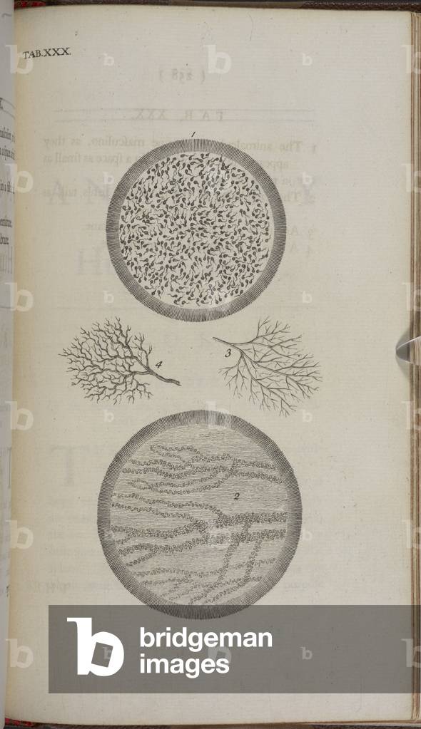
BL3314052 [The anatomy of the human body ... The fourth edition. With the addition of an appendix, etc.]Author: Chesleden, Willliam / London: W. Bowyer, 1740.Source/Shelfmark: G.19234 Tab.XXX
DC
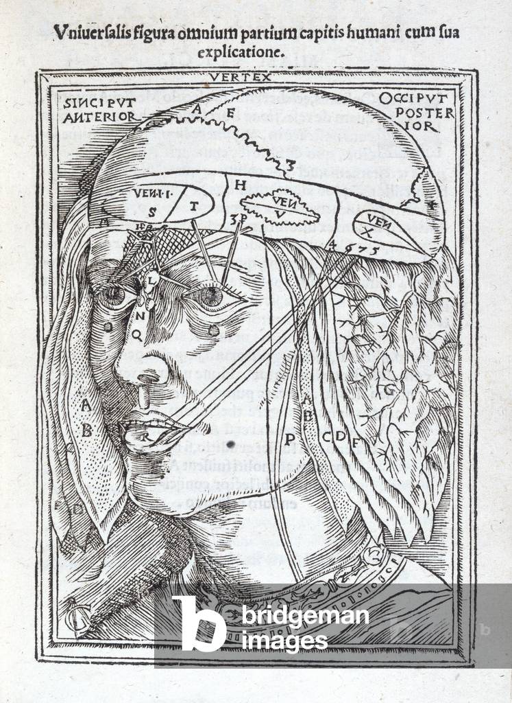
BL3265668 Anatomi: , hoc est corporis humani dissectionis parAuthor: Dryander, Johann / Marpurgi, 1537Source/Shelfmark: 548.e.29.(1), G.4 rDiagram of the human head.Image taken from Anatomi: , hoc est corporis humani dissectionis pars prior, in qua singula qu: ad caput spectant recensentur membra, atque singul: partes, singulis suis ad vivum commodissime expressis figuris, delineantur.Item Anatomia porci, ex traditione Cophonis; anatomia infantis, ex G. de Zerbis.Originally published/produced in Marpurgi, 1537.
DC
UDK844838
DC
PIX4650408 Teleportation - Artist's View
DC
FLO4591008 Anatomy of the human skeleton. Copperplate engraving by Milton from Abraham Rees' Cyclopedia or Universal Dictionary of Arts, Sciences and Literature, Longman, Hurst, Rees, Orme and Brown, London, 1820.
DC
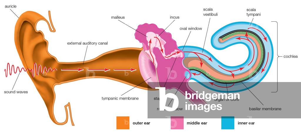
FLO4591705 Anatomy of the human ear showing ear flap, anvil, stirrrup, cochlea, eustachian tube and internal parts. Copperplate engraving by Milton from Abraham Rees' Cyclopedia or Universal Dictionary of Arts, Sciences and Literature, Longman, Hurst, Rees, Orme and Brown, London, 1820.
DC
PIX4578335 Gardening by the Moon - Michel carries the Full Moon in a barrow
DC
PIX4567079 Amateur Astronomer - Amateur Astronomer - An amateur astronomer takes a picture through a telescope. An amateur astronomer takes an astrophotograph through a telescope
DC
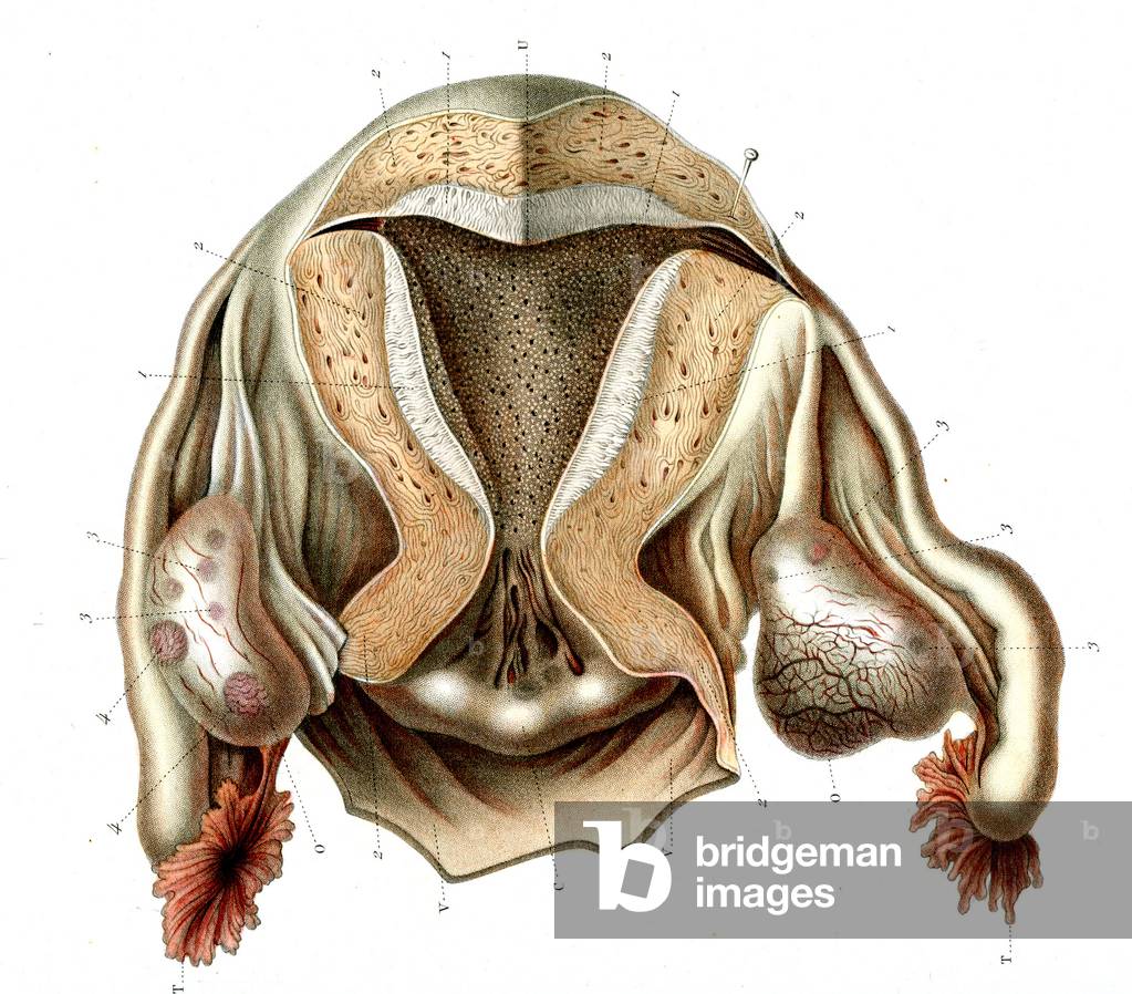
JAB4119157 Female reproductive system 1866 illustration showing the female internal reproductive organs: vagina (bottom centre), cervix (opening of the uterus, lower centre), uterus (womb, upper centre), ovaries (oval, left and right), fallopian tubes (vertical, left and right), fimbriae (feathery) of the infundibulum (far left and right) Structure of uterus, mucous uterine - Open maiden uterus, posterietric part, 1866
DC
JAB4119175 Female reproductive system 1866 illustration showing the female internal reproductive organs Fig 1: vagina (bottom centre), cervix (opening of the uterus, lower centre), uterus (womb, upper centre), ovaries (beige, oval left and right), fallopian tubes (horizontal, left and right), fimbriae (feathery) of the infundibulum (far left and right) Fig 2: vagina (vagina ( Bottom half), cervix (center), uterus (top centre) Internal genitals of women - Fig 1: vagina open forward and ovaries, Fig 2: vagina and uterus open (posterieure part), 1866
DC
JAB4119182 Virgin and non-virgin vulva anatomy 1866 illustration comparing the vulva (external reproductive organs) of a virgin (left) and non-virgin (right) female In a virgin, the hymen (12) is intact, whereas it is broken in the non-virgin The vulva consist of the labia majora, mons pubis, labia minora, clitoris, bulb of vestibule, vulval vestibule, greater and lesser vestibular acorns, external urethral orifice and the opening of the vagina (introitus) The hymen is a membrane that surrounds or partially covers the external vaginal opening (lower centre) and is typically torn during sexual intercourse External genital organs of women - Fig 1: Vulva of virgin maiden, Fig 2: Vulva de femme defloree, 1866
DC
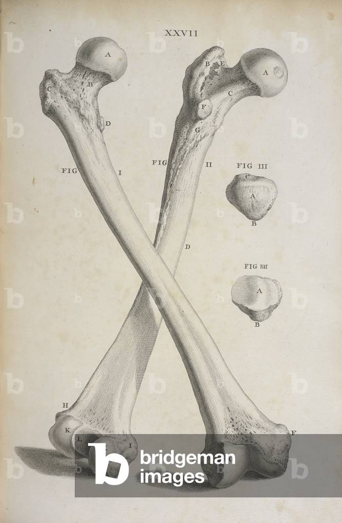
BL3314066 Osteographia, or the Anatomy of the bones.Author: Cheselden, William / London, 1733Source/Shelfmark: 458.g.1 plate XXVII
DC
FLO4594159 Anatomy of the human heart. Copperplate engraving by Milton from Abraham Rees' Cyclopedia or Universal Dictionary of Arts, Sciences and Literature, Longman, Hurst, Rees, Orme and Brown, London, 1820.
DC
UIG534867
DC
PIX4567380 Sun observation - Sun observation - Sun observation with an eclipse aim. Sun observation with a Hand held solar viewer
DC
UDK844710
DC
FLO4605235 Young Nubian woman and Ababde man. Ethiopian mixed-race people. Handcoloured steel engraving by Lizars after an illustration by Charles Hamilton Smith from his Natural History of the Human Species.
DC
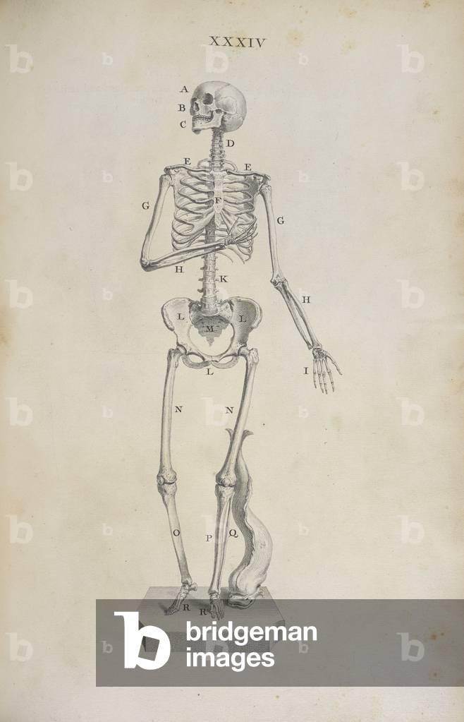
BL3314068 Osteographia, or the Anatomy of the bones.Author: Cheselden, William / London, 1733Source/Shelfmark: 458.g.1 plate XXXIV
DC
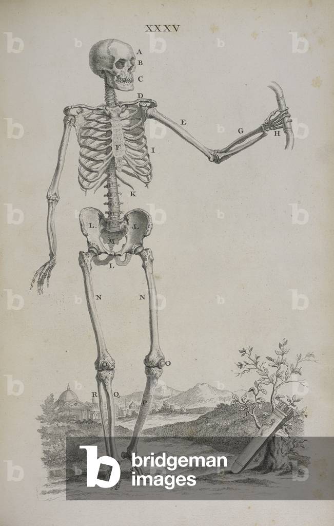
BL3314069 Osteographia, or the Anatomy of the bones.Author: Cheselden, William / London, 1733Source/Shelfmark: 458.g.1 plate XXXV
DC
PIX4567369 Sun observation with protective glasses - Sun observation with safe solar glasse
DC
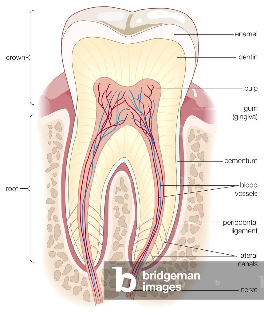
UIG535384 Cross section of an adult human molar.
DC
XJF143291 By Andreas Vesalius
RF
UIG533863
DC
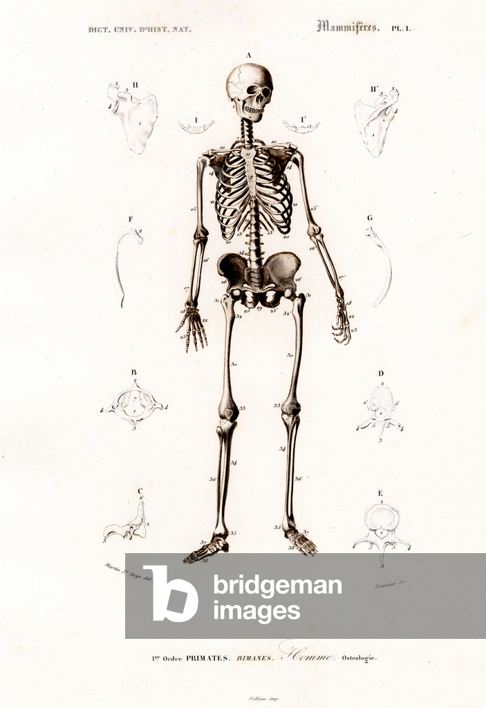
PRX11859126 This detailed anatomical illustration shows a complete human skeleton in frontal view, surrounded by specific bone details of individual body regions. The image is labeled with letters and numbers that identify anatomical structures for didactic purposes. By Martin St. Ange del., Fournier sc. From Dictionnaire Universel d'Histoire Naturelle dirigé par M. Charles D'Orbigny. Paris, chez les Éditeurs MM. Renard, Martinet et Cie, 1849. [anatomy, osteology, human skeleton, bone structure, primates, Homo sapiens, skull, spine, pelvis, extremities, natural history, 19th century, scientific illustration, mammals, hand-colored steel engraving]
DC
UDK844268
DC
UDK844267
DC
UDK844270
DC
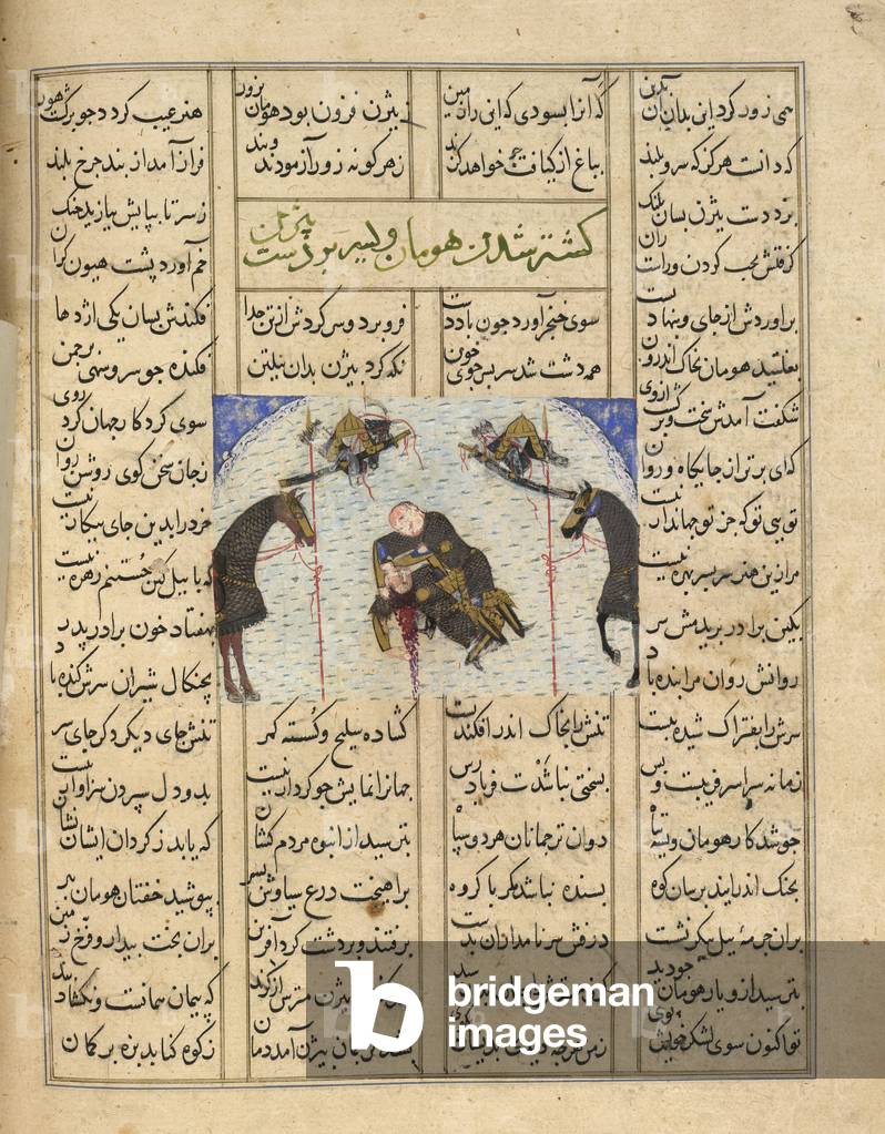
BL3264937 Shahnama.Author: Firdawsi / Iran, 1446Language: PersianSource/Shelfmark: Or. 12688, f.283vBizhan killing Human. A miniature painting from a fifteenth century manuscript of the epic poem of Shahnama.Image taken from Shahnama.Originally published/produced in Iran, 1446.
DC

Total de Resultados: 62.869

Página 1 de 629